


SKU:
IBOOLO
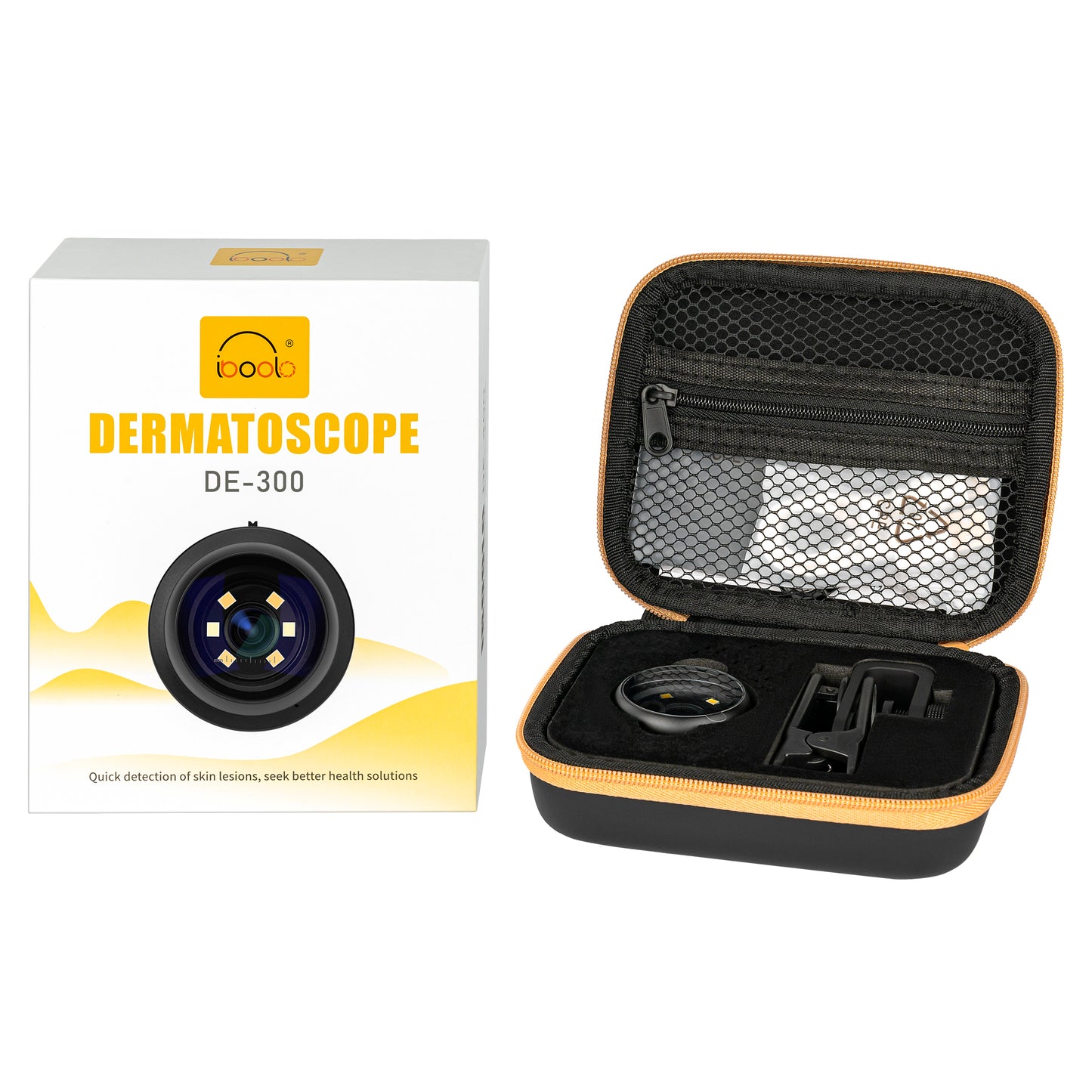
IBOOLO DE-300 Economic Dermatoscope Skin Analyzer Machine for Primary Care
IBOOLO DE-300 Economic Dermatoscope Skin Analyzer Machine for Primary Care
Used by Dermatologist in 30+countries
FDA-Cleared CE Certified
Tilgængelighed for afhentning kunne ikke indlæses
Advantages of DE-300
The IBOOLO DE-300 dermoscope is ideal for home use and hospital use. Combined with a smartphone, you can get amazing, high-quality images. It contains a universal phone adapter meaning convenient to connect with any smartphone or tablet to capture images.
• Polarized and non-polarized lighting
• Real 6x Magnification
 Technical Specifications
![]()
| Name | Dermatoscope/Dermoscopy |
|
Item |
DE-300 |
| Material | Optical and Aluminum |
| Magnification | 6 times |
| LEDS | 6 LEDS |
| Light | Polarization and non polarization |
| Warranty | 2 Years |
| Certificate | CE FDA |
| Standard | ISO13485 ISO14001 |
| Brightness | 1 |
| Battery | 200mAh |
DE-300 Dermatoscope Features
This top-tier dermatoscope combines exceptional performance with affordability, making it an ideal choice for primary dermatology care. It features both polarized and non-polarized light modes, enabling comprehensive skin examinations by minimizing surface reflections and revealing deeper skin structures. Its user-friendly design and reliable functionality ensure accurate diagnostics, catering to a wide range of dermatological needs.

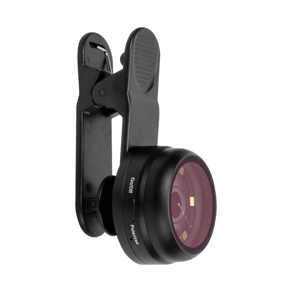
Lighting System
The DE-300 dermatoscope can be switched to both polarized and non polarized light model, available in different lesion diagnose situation.
The non polarized light is mainly used to observe superficial skin lesions with the help of visible light;
The polarized light can help to eliminate surface reflection and allow visualization of vascular structures. The instrument allows for visualization for deeper skin structures, such as blood vessels, collagen, and pigment in the dermis.

Compatibility
IBOOLO DE-300 Dermatoscope Camera for Smartphone Skin Imaging
The IBOOLO DE-300 is a portable dermatoscope camera designed for smartphone dermoscopy imaging. With 6x magnification, polarized and non-polarized LED lighting, a rechargeable battery, and a universal phone adapter, it supports close-up skin visualization, image capture, and clinical documentation for primary care, dermatology, education, and mobile screening workflows.
IBOOLO DE-300 Dermatoscope Camera Features
- 6x optical magnification: Supports close-up visualization of skin surface details.
- Polarized and non-polarized lighting: Allows users to switch between different dermoscopy viewing modes.
- Smartphone and tablet compatibility: Includes a universal phone adapter for mobile dermoscopy image capture.
- Rechargeable design: Built-in battery with USB-C charging for portable clinical use.
- Portable entry-level design: Suitable for primary care, medical education, dermatology clinics, and mobile examination workflows.
DE-300 Dermatoscope Technical Specifications
| Product Name | IBOOLO DE-300 Dermatoscope Camera |
|---|---|
| Product Type | Portable smartphone-compatible dermatoscope |
| Magnification | 6x |
| Lighting | Polarized and non-polarized LED illumination |
| LEDs | 6 LEDs |
| Compatibility | Universal phone adapter for smartphones and tablets |
| Battery | Rechargeable 200mAh battery |
| Warranty | 2 years |
Smartphone Dermoscopy Camera for Skin Imaging
The DE-300 works as a smartphone-compatible dermoscopy camera when used with the included universal phone adapter. It helps users capture close-up dermoscopic images with a smartphone or tablet, making it suitable for documentation, follow-up comparison, patient communication, and teaching purposes.
Unlike a basic optical dermatoscope that only supports direct visual examination, the DE-300 supports mobile image capture through compatible devices. This makes it a practical option for clinics, primary care practices, medical students, and users who need a portable dermatoscope camera without a large digital imaging system.
Looking for a Firefly DE300 Alternative?
For users comparing Firefly DE300-style dermatoscope cameras, the IBOOLO DE-300 offers a portable and cost-effective alternative for smartphone dermoscopy. It combines 6x magnification, polarized and non-polarized LED lighting, and a universal phone adapter for mobile image capture.
| Comparison Point | IBOOLO DE-300 | Firefly DE300-style Search Intent |
|---|---|---|
| Product Type | Portable smartphone-compatible dermatoscope | Digital dermatoscope camera / alternative device |
| Lighting | Polarized and non-polarized LED light | Users usually compare illumination and imaging clarity |
| Image Capture | Via smartphone or tablet adapter | Users want digital documentation capability |
| Best Fit | Primary care, education, mobile checks, clinic documentation | Users looking for a portable dermatoscope camera alternative |
Frequently Asked Questions About the DE-300 Dermatoscope
Is the IBOOLO DE-300 a dermatoscope camera?
Yes. The IBOOLO DE-300 can function as a dermatoscope camera when used with the included universal phone adapter. It allows users to capture dermoscopic images through a compatible smartphone or tablet.
Is the DE-300 a digital dermatoscope?
The DE-300 is best described as a smartphone-compatible digital dermoscopy device. It is not a large standalone digital dermatoscope system, but it supports digital image capture when connected with a smartphone or tablet.
What is the difference between DE-300 and a traditional dermatoscope?
A traditional dermatoscope is mainly used for direct visual examination. The DE-300 adds smartphone and tablet compatibility, allowing users to capture and document skin images for review, follow-up comparison, and teaching.
Can the DE-300 be used as a Firefly DE300 alternative?
Yes, users searching for a Firefly DE300 alternative may consider the IBOOLO DE-300 as a portable smartphone-compatible dermatoscope option. It is not a Firefly product, but it provides a similar portable dermoscopy camera use case.
Who is the DE-300 suitable for?
The DE-300 is suitable for primary care providers, dermatology clinics, medical students, training settings, and users who need a portable dermatoscope camera for close-up skin imaging and documentation.
Package Include
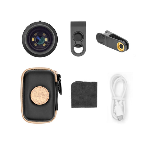

- DE-300 Dermatoscope
- Universal Phone Adapter
- Clean Cloth
- USB-C Cable
- Carry Case
- English Version User Manaul


DE-4100/4100 PRO Gallery


